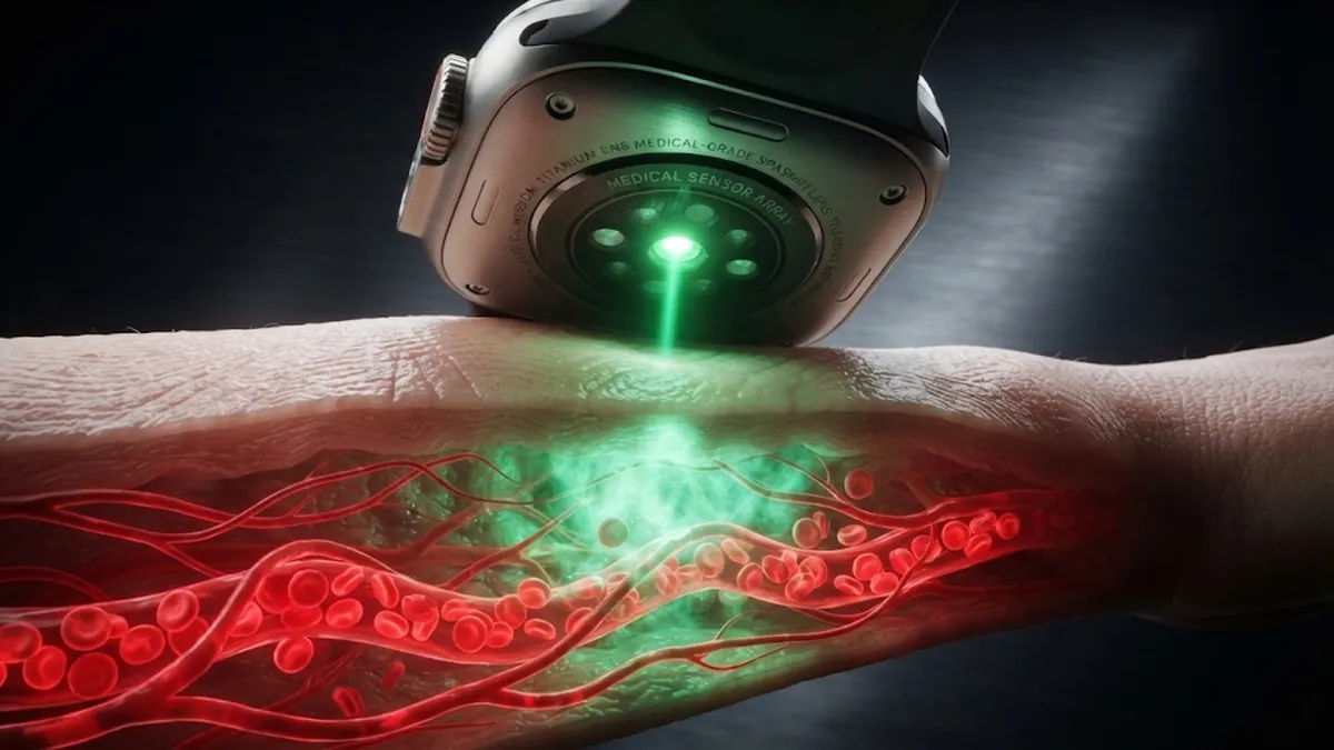

Flashing Green in the Dark: The Optics Beneath the Sapphire Crystal
If you take off your smartwatch in a dim bedroom, you will catch a cluster of tiny emerald-green lights strobing against the nightstand. Those lights are not indicator signals. They are high-speed optical emitters firing hundreds of times every second, driving beams of light straight through your epidermis to measure physical blood volume. Here is how photoplethysmography (PPG) turns light reflections into accurate heart rate readings without a single needle or electrical lead.
1. The Underside Sensor Cluster: LEDs and Photodiodes
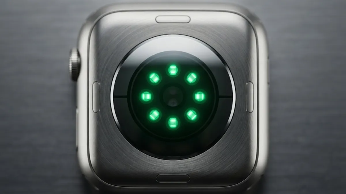
Every modern wearable health monitor relies on a pair of complementary optical hardware components: light-emitting diodes (LEDs) and silicon photodiodes. The LEDs act as transmitters, showering the tissue with photons at calibrated wavelengths. The photodiode sits in the center as the receiver, converting returning light intensity into tiny micro-ampere electrical currents. As arterial blood pulses through your wrist with each contraction of the heart, the volume of blood under the sensor expands and contracts rhythmically.
2. Why Green? The Hemoglobin Absorption Secret
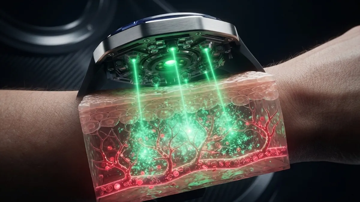
Blood is red because it reflects red light and absorbs green light. In the visible spectrum, oxygenated hemoglobin has an exceptionally high optical absorption coefficient at approximately 525 to 535 nanometers (green light). When your heart beats, blood surges through the arterioles in your wrist, absorbing more green photons and reducing the amount of light bounced back to the photodiode. Between beats, when blood volume recedes, more green light reflects back. By measuring these peak-and-trough absorption cycles hundreds of times per second, the processor calculates your exact pulse rate.
3. The Motion Problem: Running, Sweat, and Light Leaks
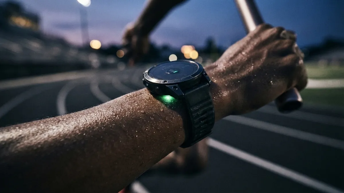
Optical PPG works flawlessly when you sit motionless at a desk. But during an all-out sprint, the watch bounces against your skin, introducing massive optical noise called motion artifacts. Ambient sunlight leaks under the watch casing, flooding the photodiode with millions of competing photons. To solve this, manufacturers use dual-wavelength subtraction and high-rate digital filtering. Built-in accelerometers measure the exact rhythm of your arm swings and mathematically subtract that motion frequency from the optical raw data stream.
4. The Ink Barrier: Why Tattoos Confuse the Sensor
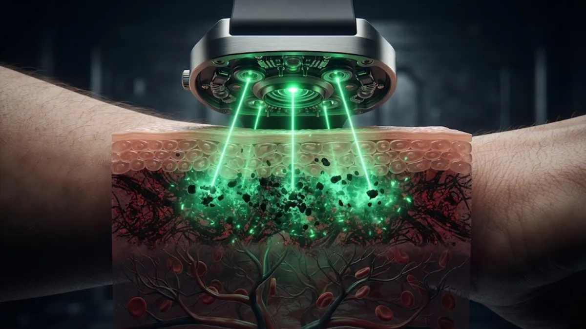
One of the most common user complaints is that optical sensors misread or stop tracking over tattooed wrists. Tattoo needles deposit insoluble pigment particles deep inside the dermis—the exact skin layer where capillary blood vessels live. Dark black and red tattoo inks contain carbon and heavy metal oxides that absorb green light aggressively, blocking the light from reaching the capillaries and preventing the photodiode from receiving returning signals. In response, modern devices dynamically increase LED drive current or fall back to deeper-penetrating infrared wavelengths.
Optical Biosensing: Sensor Wavelengths Compared
| Wavelength Band | Light Spectrum | Skin Penetration Depth | Primary Diagnostic Use |
|---|---|---|---|
| 525 – 535 nm | Emerald Green | Shallow (Dermis / Capillary loops) | Continuous pulse tracking, athletic heart rate |
| 660 nm | Deep Red | Medium (Subdermal microvessels) | SpO2 blood oxygen saturation calculations |
| 940 nm | Near-Infrared (NIR) | Deep (Arterioles and deep tissue) | Resting pulse rate, low-power background health checks |
Engineering Takeaway: Why Not Just Use Infrared All Day?
Infrared light penetrates deeper into human tissue and consumes significantly less battery power. However, infrared is vulnerable to motion artifacts during physical exercise. Green light has a far superior signal-to-noise ratio in active capillary beds, which is why your watch switches aggressively to pulsing green whenever you start moving.