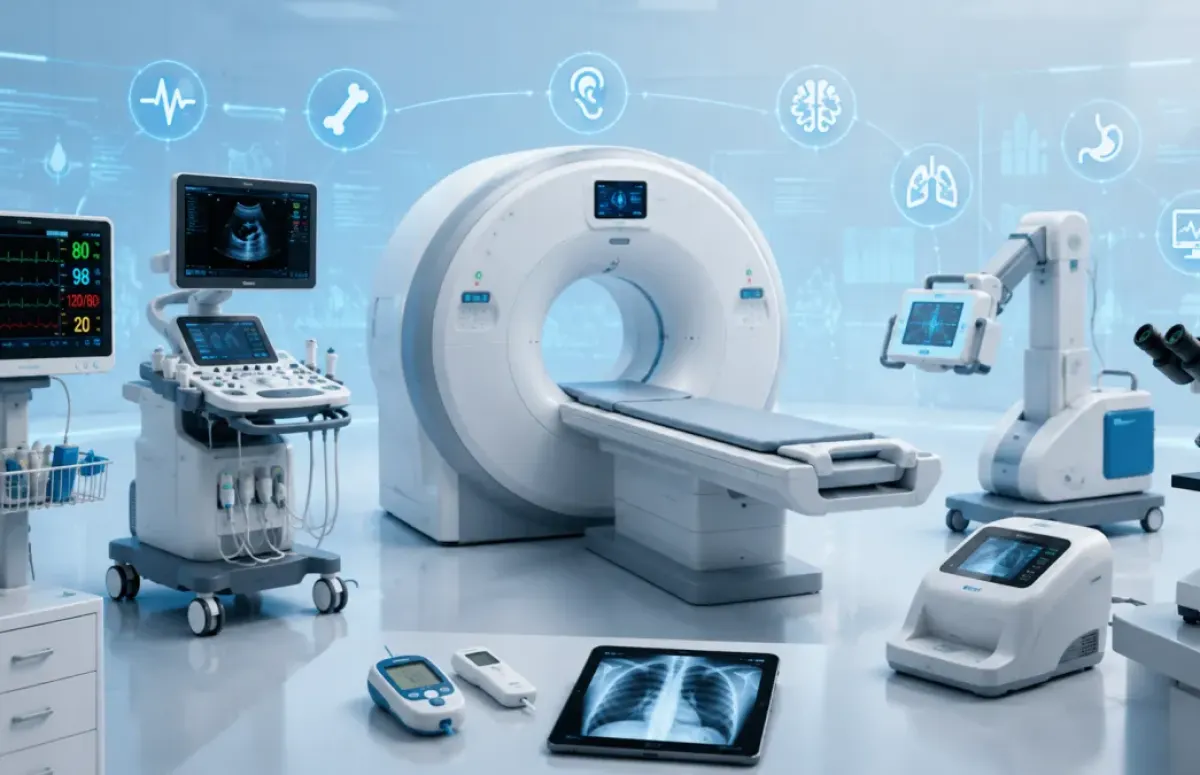
Modern medicine depends heavily on advanced diagnostic tools that help doctors detect diseases earlier, faster, and more accurately. From imaging technologies to portable monitoring devices, these medical instruments play a critical role in improving patient outcomes and saving lives. In this guide, we explore 15 of the most important medical diagnostic devices and explain how they work, their key applications, and why they matter in healthcare today.
1. MRI Scanner (Magnetic Resonance Imaging)
An MRI scanner uses a powerful magnetic field — usually between 1.5 and 3 Tesla, and up to 7 Tesla in advanced systems — along with radio waves to create detailed images of soft tissues inside the body. Unlike CT scans, MRI does not use ionizing radiation.
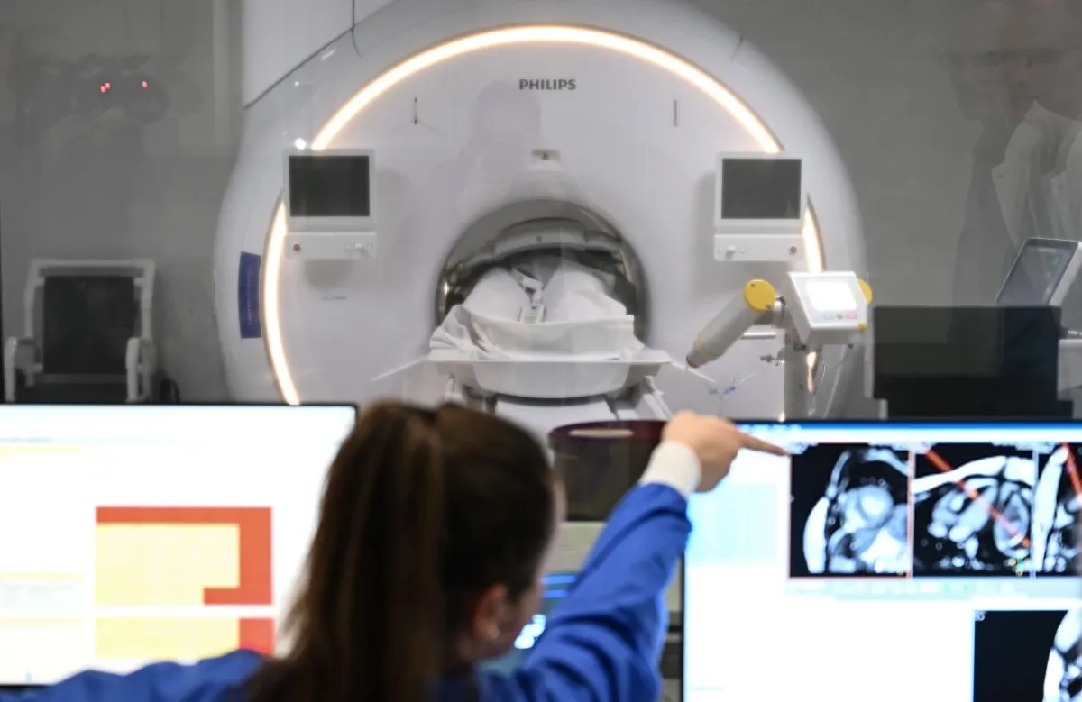
MRI works by aligning hydrogen atoms in the body with a magnetic field and then stimulating them with radiofrequency pulses. It is highly effective for diagnosing brain tumors, multiple sclerosis (MS), spinal cord injuries, ligament tears, and joint disorders.
Important note: Patients with older pacemakers or certain metal implants may not be eligible for MRI scans.
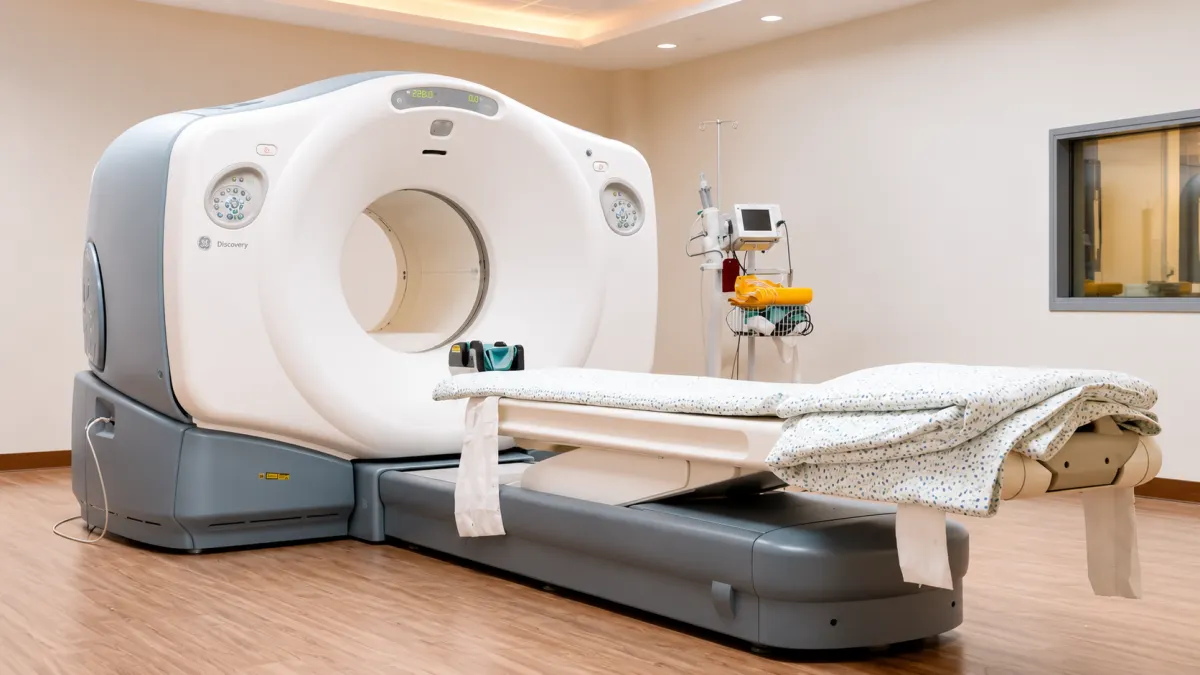
2. CT Scan (Computed Tomography)
A CT scan combines X-ray technology with computer processing to create cross-sectional images of the body. The scanner rotates around the patient and captures images from multiple angles.
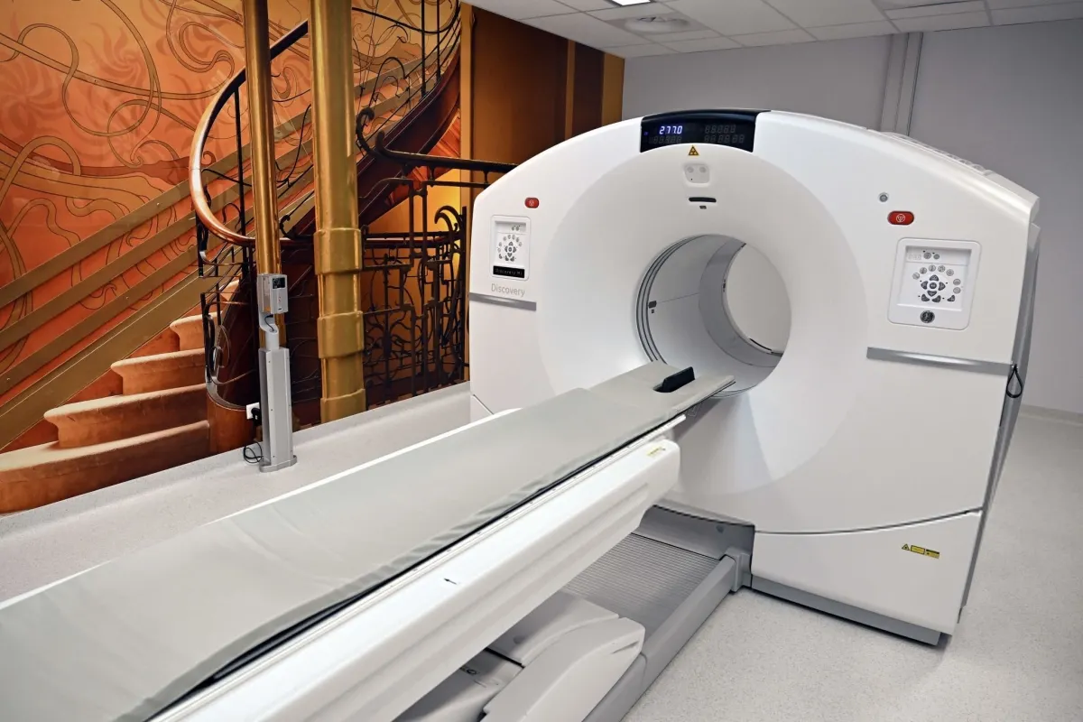
Because of its speed, CT scanning is essential in emergency medicine for detecting strokes, internal bleeding, lung embolisms, and traumatic injuries.
Educational fact: A chest CT scan exposes patients to approximately 70–100 times more radiation than a standard chest X-ray, which is why it is only used when medically necessary.
Artificial Intelligence in General Practice: Replacement, Augmentation, or a New Standard of Care?
3. PET Scan (Positron Emission Tomography)
A PET scan measures metabolic activity rather than just body structure. Patients receive a small amount of radioactive tracer, commonly fluorodeoxyglucose (FDG), which is absorbed more by active cells such as cancer cells.
PET scans are widely used in cancer staging, detecting metastasis, and evaluating neurological diseases like Alzheimer’s disease. PET is often combined with CT imaging in a PET-CT system to provide both structural and functional information.
4. Ultrasound Machine
Ultrasound imaging uses high-frequency sound waves, typically between 2 and 18 MHz, to create images of internal organs and tissues.
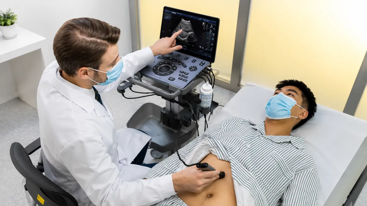
It is radiation-free, portable, affordable, and safe, making it the gold standard for pregnancy monitoring. Doppler ultrasound can also measure blood flow and help diagnose blood clots (DVT), narrowed arteries, and heart conditions.
Limitation: Sound waves do not travel well through bone or air, so ultrasound is less effective for examining lungs and the adult brain.
5. Electrocardiogram (ECG or EKG)
An ECG records the electrical activity of the heart using electrodes attached to the skin. A standard ECG provides 12 different views of heart activity.
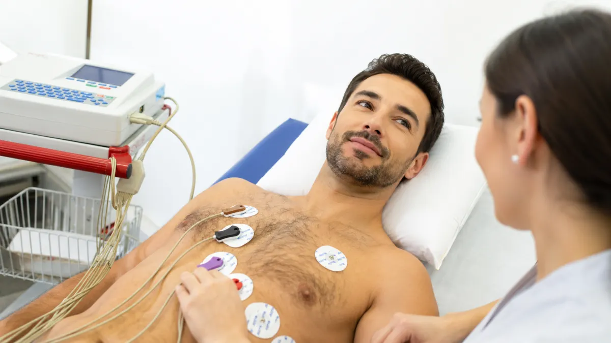
It can quickly detect heart attacks, arrhythmias, enlarged heart chambers, and electrolyte imbalances.
Key ECG Components:
- P wave: Atrial contraction
- QRS complex: Ventricular contraction
- T wave: Ventricular recovery phase
Changes in the ST segment are critical for diagnosing acute heart attacks.
6. Holter Monitor
A Holter monitor is a portable ECG device that continuously records heart activity for 24–48 hours or longer.
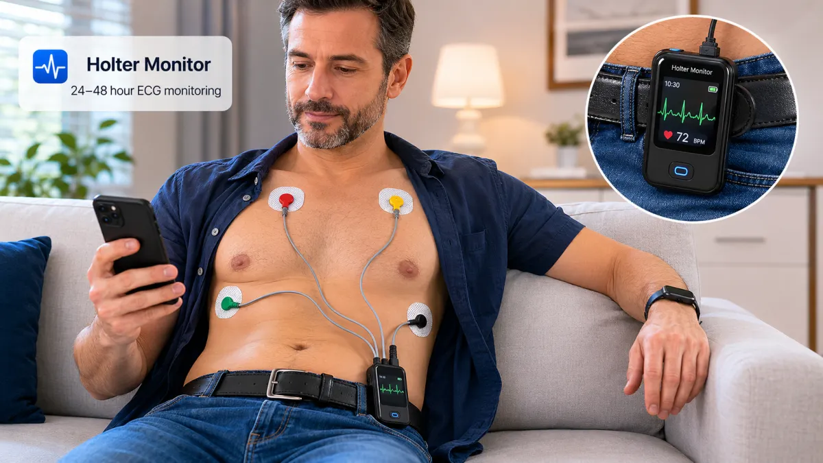
It is especially useful for patients with symptoms like palpitations, dizziness, or fainting that may not appear during a regular ECG performed in a clinic.
7. EEG (Electroencephalogram)
An EEG records electrical activity in the brain using electrodes placed on the scalp.
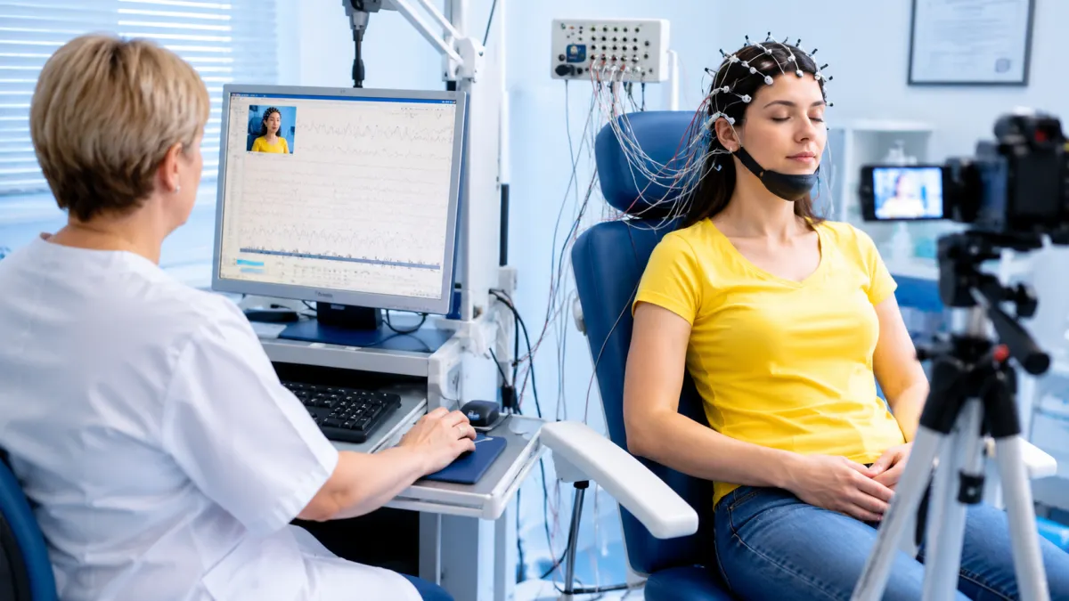
It is the primary diagnostic tool for epilepsy because it can detect abnormal brain wave patterns. EEG is also used for sleep disorders, encephalitis, and confirming brain death.
Brain waves are classified into delta, theta, alpha, beta, and gamma waves, each associated with different states of brain activity.
8. Echocardiography (Echocardiogram)
An echocardiogram is a specialized ultrasound of the heart that evaluates heart valves, chamber size, wall thickness, and ejection fraction (EF).
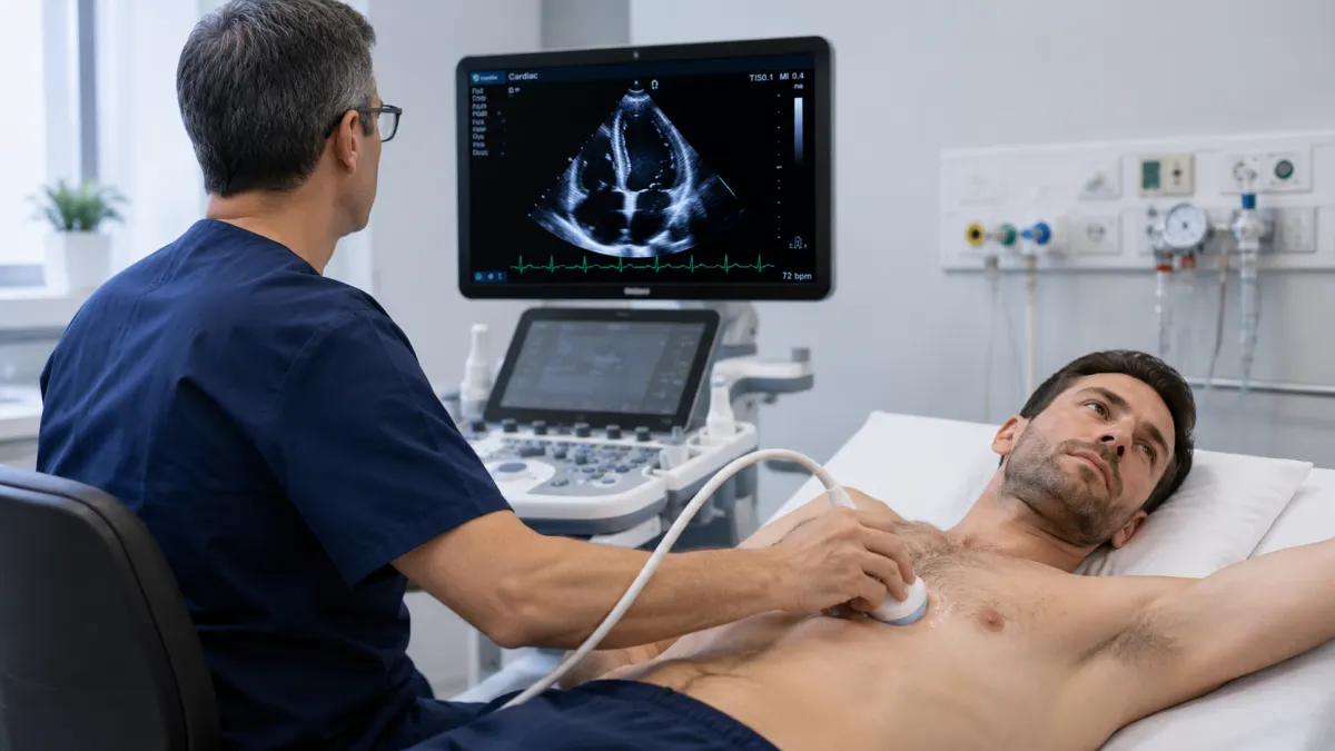
A normal EF ranges between 55% and 70%, while an EF below 40% may indicate heart failure.
A transesophageal echocardiogram (TEE), performed through the esophagus, provides more detailed images of heart valves and the left atrium.
9. Endoscopy and Colonoscopy
These procedures use a flexible tube equipped with a camera and light source.
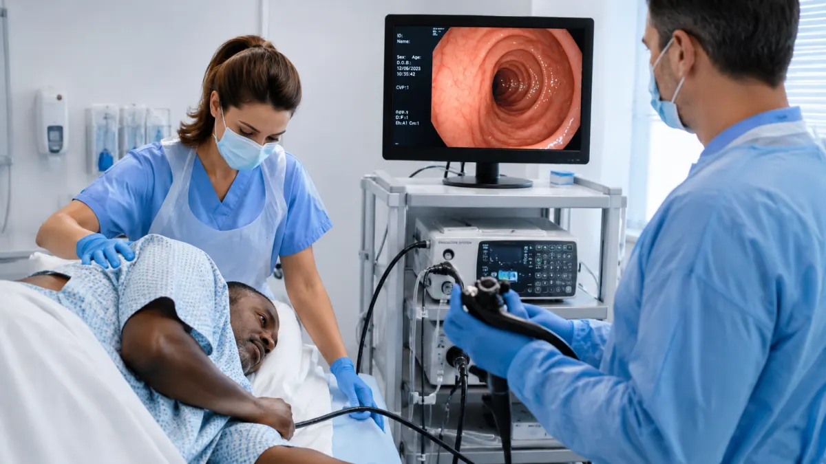
- Endoscopy: Inserted through the mouth to examine the digestive tract
- Colonoscopy: Inserted through the rectum to examine the colon
In addition to visual inspection, doctors can perform biopsies or remove polyps during the same procedure.
Colonoscopy remains the gold standard for colorectal cancer screening and is generally recommended every 10 years starting between ages 45 and 50.
10. Mammography
Mammography is a specialized low-dose X-ray imaging technique used for early breast cancer detection.
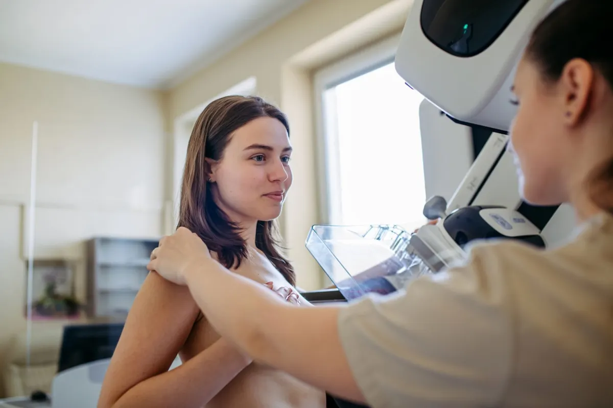
It can identify microcalcifications — tiny calcium deposits that may be one of the earliest signs of breast cancer — years before a lump becomes detectable.
Modern 3D mammography (tomosynthesis) improves accuracy, especially in women with dense breast tissue.
11. Pulse Oximeter
A pulse oximeter is a small device placed on a fingertip to measure blood oxygen saturation (SpO2).
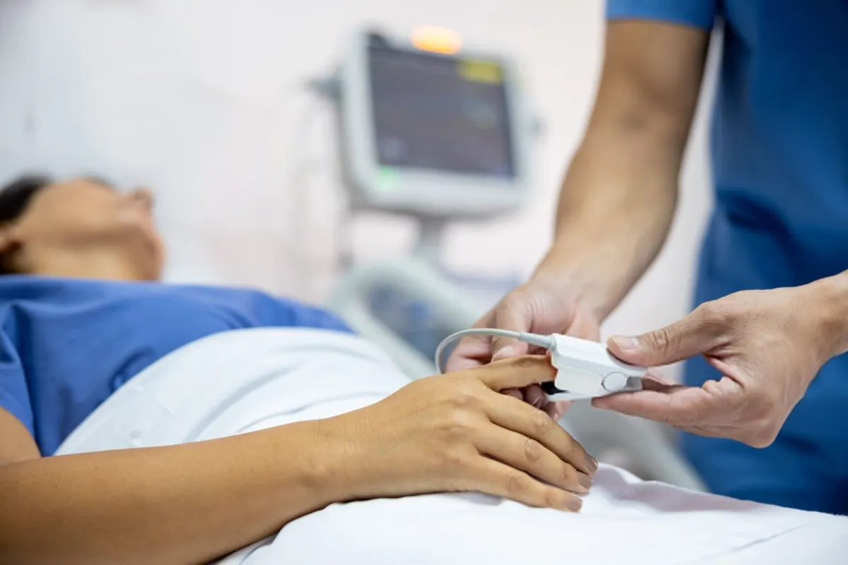
It works by passing red and infrared light through the finger. Normal oxygen saturation levels range from 95% to 100%.
Pulse oximeters became globally important during the COVID-19 pandemic because they helped identify silent hypoxia — dangerously low oxygen levels without obvious symptoms.
Important note: Nail polish, cold hands, and poor circulation may affect accuracy.
12. Spirometer
A spirometer measures lung function by analyzing airflow and lung capacity during breathing.
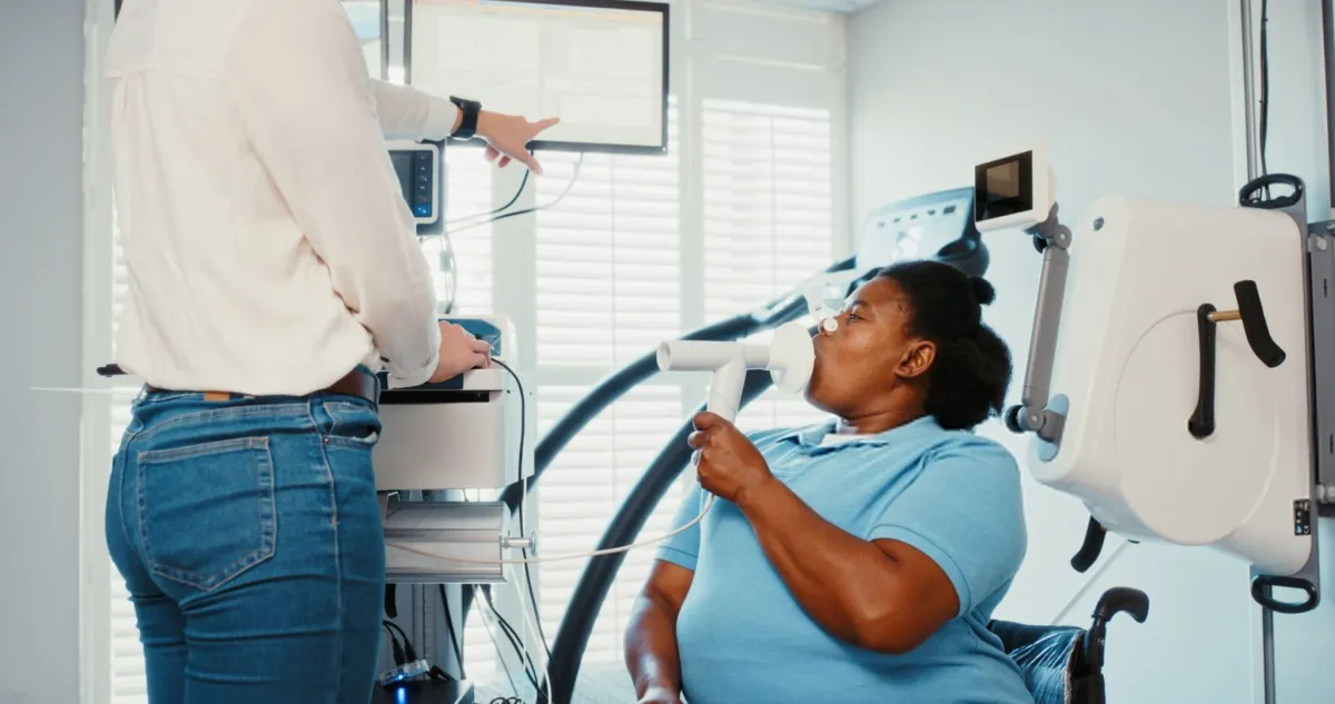
Two key measurements include:
- FEV1: Forced expiratory volume in one second
- FVC: Forced vital capacity
An FEV1/FVC ratio below 0.7 often indicates obstructive lung diseases such as asthma or COPD.
Spirometry is essential for diagnosing respiratory diseases, determining severity, and monitoring treatment response.
13. Glucometer and Continuous Glucose Monitoring (CGM)
A traditional glucometer measures blood sugar using a small drop of blood from the fingertip.
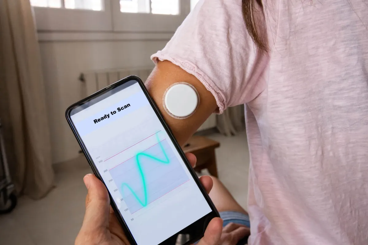
Newer Continuous Glucose Monitoring (CGM) systems use a sensor placed under the skin to track glucose levels continuously and send data to a smartphone or insulin pump.
CGM technology has significantly improved diabetes management, especially for patients with Type 1 diabetes.
14. Otoscope and Ophthalmoscope
These are simple but essential examination tools used in everyday clinical practice.
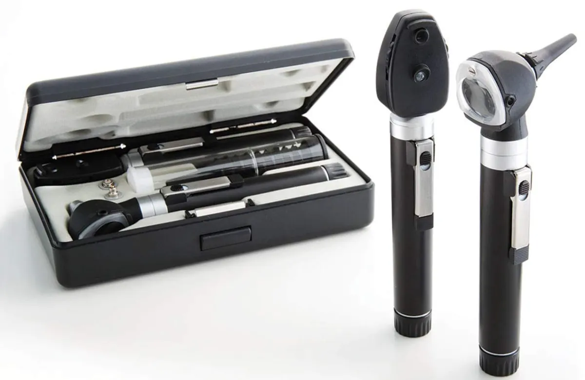
- Otoscope: Used to examine the ear canal and eardrum for infections, fluid buildup, or perforation
- Ophthalmoscope: Used to examine the retina and optic nerve
Eye examinations can help detect diabetic retinopathy, glaucoma, papilledema, and even high blood pressure because retinal blood vessels can be directly observed.
15. DEXA Scan (Bone Density Scan)
A DEXA scan uses two low-dose X-ray beams to measure bone mineral density, especially in the spine and hip.
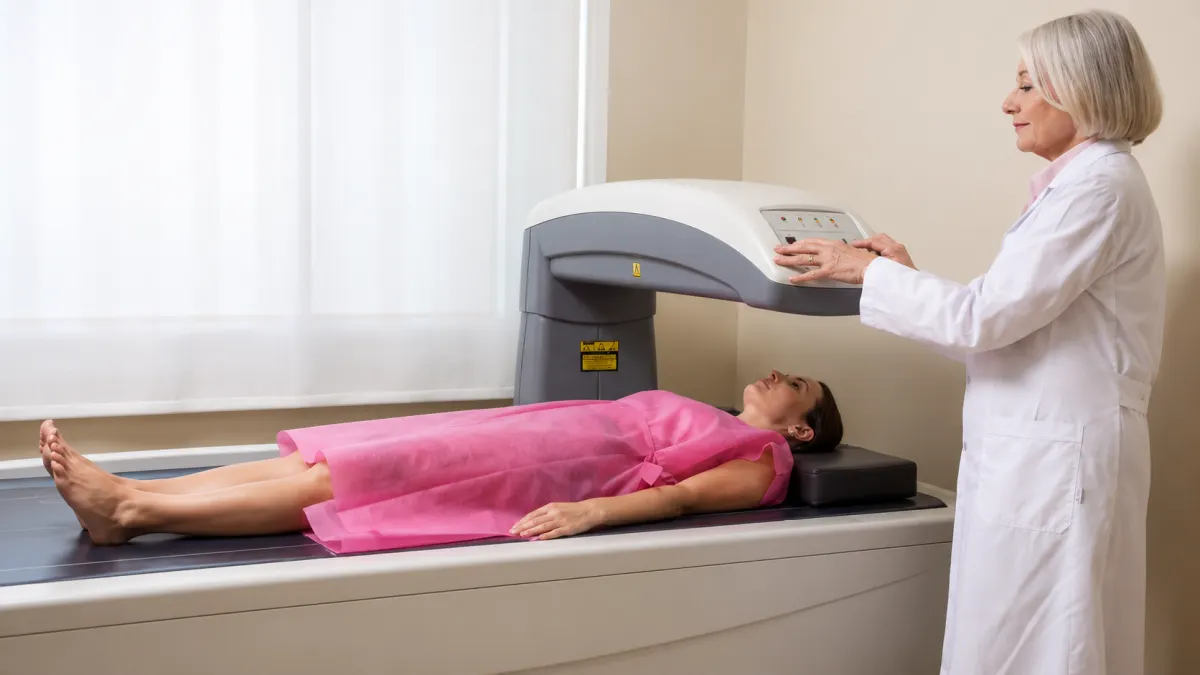
Results are reported using a T-score:
- Above -1: Normal
- Between -1 and -2.5: Osteopenia
- Below -2.5: Osteoporosis
Early detection of osteoporosis is particularly important in postmenopausal women to help prevent fractures.